Cell Biology – Cell Culture Quality Control Assays
September 2023
Automated imaging tools can provide researchers with valuable information for improving routine cell culturing techniques and increasing the effectiveness and reproducibility of downstream cell-based assays. Conventional methods for evaluating cell culturing techniques and assay optimization consist of manual inspection of a small subset of the cell population at random locations and time points. These methods rely on subjective measurements that are difficult to standardize and contribute to increased variability from round to round. In contrast, continuous monitoring of cell cultures using instruments capable of automated image capture and analysis provides a detailed record of a broad range of cell characteristics.
Agilent BioTek instruments, including the Lionheart FX and Cytation automated imaging systems, MultiFlo FX multimode dispenser, and the BioSpa live cell analysis system are designed to improve the accuracy and reproducibility of cell-based assays from culture to analysis.
In this edition of TekTalk, we have compiled diverse resources describing how automated imaging tools and analysis can be employed to conduct automated cell culture quality control studies and assay optimization. Content includes tips and techniques for monitoring cell growth characteristics, measuring cell culture density within labware, quantifying transduction/transfection efficiency, and more.
Featured Applications
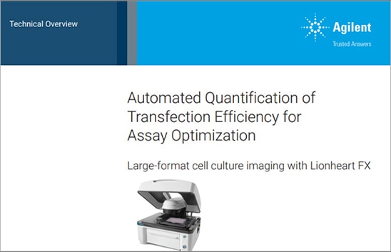
Automated Quantification of Transfection Efficiency for Assay Optimization
Chemical transfection is a well-established approach to introduce plasmid DNA for gene expression into mammalian host cells. The transfection efficiency (TE) of a given plasmid is quantified by deriving a ratio between the number of transfection cells and the total number of cells counted. This is typically accomplished using fluorescent reporters and a microscope, with one or more image fields acquired within the culture vessel where the transfection took place.
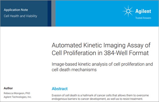
Automated Kinetic Imaging Assay of Cell Proliferation in 384-Well Format
Evasion of cell death is a hallmark of cancer cells that allows them to overcome endogenous barriers to cancer development, as well as to resist treatment. Understanding how cancer cells are able to evade cell death promises to unlock new avenues to treat human cancer. This application note presents a fully automated image-based assay to monitor both the proliferation and cell death of a fibrosarcoma cancer cell line in response to multiple antineoplastic drugs in a high-throughput format.
Application Compendium
Automated Methods for Conducting Cell Culture Quality Control Studies and Assay Optimization
Automated imaging tools can provide researchers with valuable information for improving routine cell culturing techniques and increasing the effectiveness and reproducibility of downstream cell-based assays. In this compendium, we describe quantitative imaging-based methods designed to provide accurate and objective evaluation of several important variables that contribute to experimental results.
Featured Product
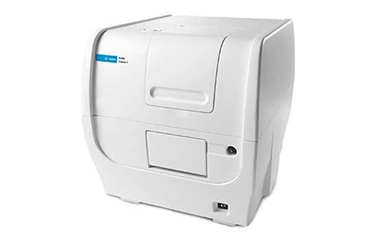
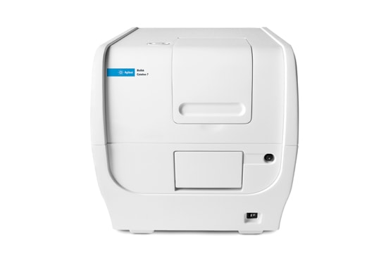
Agilent BioTek Cytation 7 Cell Imaging Multimode Reader
The BioTek Cytation 7 cell imaging multimode reader combines automated digital upright and inverted widefield microscopy with monochromator-based multimode microplate reading. The inverted microscope provides 1.25x to 60x magnification in fluorescence, brightfield and color brightfield, while the upright microscope enables other common applications including ELISpot, slide scanning and ROI detection.
Tek Tips
Technique for achieving an even distribution of cells within microplate wells
One common lab practice contributing to uneven distribution of cells within microplate culture vessels is placing the microplate directly into the culture incubator immediately after seeding. The rapid change in temperature promotes a higher density of cells at the perimeter of the culture chamber compared to the center. To reduce this effect, consider keeping microplate culture vessels at room temperature within the environment hood after seeding for approximately 10-30 minutes, depending on culture volume and cell characteristics. This step allows adherent cells to evenly settle down and attach to the culture surface before transferring the vessel to a cell culture incubator.
Optimize staining protocols for kinetic live cell applications
A broad range of fluorescent stains are available for live cell studies. However, the addition of these reagents can produce unintended cell type-dependent cytotoxic effects. Agilent BioTek Gen5 image analysis tools support the ability to determine the appropriate concentrations of reagents to achieve efficient labeling throughout the experiment without significantly altering cellular behavior in the process. The minimal amount of stain necessary to effectively identify cells should be determined by imaging the cells across a reasonable range of stain concentrations and determining the signal-to-noise ratios over the intended length of the experiment. Note, cells will often exhibit a decrease in stain intensity over time within culture. In parallel, test for stain-induced effects on cell health and proliferation resulting from each stain concentration using the Automated Kinetic Imaging Assay of Cell Proliferation assay described in the application note above. Proceed with a stain concentration that supports robust identification of cells throughout the time course while having minimal impact on cell behavior.
Resources
Application Compendium
Application Notes
- Automated Media Exchanges Preserves 3D Samples for Long-Term Maintenance
- Image-based label-free methods to detect and quantify General and Directed T-cell Activation
Research Article
Technical Overview
Webinars on Demand
Tools and Techniques for Optimizing Cell Proliferation Studies
Presenter: Joseph E. Clayton, Ph.D., Global Scientific Program Manager, Agilent
In this webinar we provide a detailed review of available methods for measuring cell proliferation, with a particular focus on recent assay innovations, including both 2D and 3D formats. This information will assist researchers in selecting the most appropriate and effective system for measuring cell proliferation based on a broad range of factors. Additionally, we cover important considerations for setting up and running cell proliferation assays, including techniques for improving the accuracy and reproducibility of results.
CAR-T: from Discovery to Process Development and Manufacturing, hosted by Agilent Cell Analysis
Presenters: Roddy O'Connor, PhD, Perelman School of Medicine, University of Pennsylvania; Joseph Lee, PhD, Affini-T Therapeutics; Tamara Laskowski, PhD, Personalized Medicine, Lonza Global
The landscape of cell and gene therapy is rapidly evolving, and researchers are seeking opportunities to bring safe and effective therapies to patients–by improving the quality and enhancing the performance of cell products and streamlining the cell biomanufacturing process. There is a critical need for biologically relevant assays to identify critical quality attributes to support cell therapy discovery research, cell engineering, reduce variability, and streamline process development and manufacturing.
Join us for a virtual symposium and panel discussion from key opinion leaders in cell and gene therapy to discover new advancements in discovery research, and learn how they overcome challenges in cellular engineering, process development and manufacturing.
Promotions
View specific promo details to determine availability in your region.
Buy a Synergy H1M or H1MF and get 35% off
Customers who purchase a Synergy H1M or Synergy H1MF configuration of the Synergy H1 model are eligible to receive 35% promotion discount (total of promotion+purchase agreement discount).
Equip your new lab with the latest Agilent Cell Analysis Technologies
Save 25% when you buy two or more Agilent Cell Analysis instruments for your new lab or startup lab.
Do You Own an Agilent BioTek Synergy Multimode Reader? This Offer is for You!
Buy an eligible Agilent BioTek Cytation cell imaging multimode reader and get 25% off!
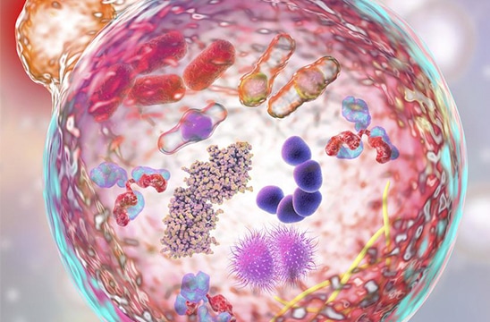
Explore Next Generation Solutions for Cell Therapies
In this virtual lab, explore solutions for cell engineering, immunophenotyping, and high-throughput screening of optimal engineered immune cells. The success of these cell therapies depends on selective engineering of immune cells followed by extensive ex-vivo characterization to identify the optimal candidate. Agilent offers a growing number of solutions to assess engineered immune cell activation, potency, exhaustion, and metabolic phenotypes to determine cell fate, fitness, and function.