Celebrating a Decade of Imaging & Microscopy
May 2023
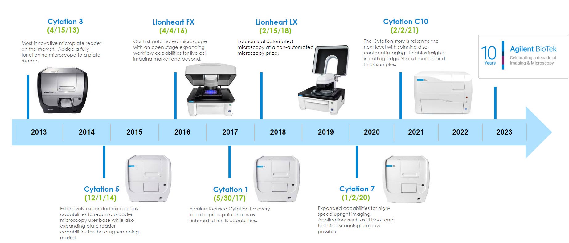
In 2013, the unique capabilities of the Cytation 3 were the first of its kind, combining both microscopy and multimode plate reading in a single instrument. This innovative benchtop solution made imaging affordable for many labs that wouldn't have been able to incorporate a typical fully automated imaging solution into their research "toolset". The Cytation product line has evolved over the last 10 years as we incorporated customer feedback to build imaging and microscopy systems that enable workflows that answer fundamental research questions in the realm of cell analysis, drug discovery and more.
Imaging Product Timeline (enlarge)
Featured Applications

Characterizing Calcium Mobilization Using Kinetic Live Cell Imaging
Ca2+ acts as an important second messenger in diverse signaling pathways, including G protein-coupled receptors. Characterizing these pathways requires the ability to detect rapid changes in intracellular Ca2+ levels with high temporal resolution. This application note describes a live cell imaging-based approach to quantify Ca2+ flux kinetics using an Agilent BioTek Lionheart FX and Fluo-4 Ca2+ indicator dye that delivers sub-second resolution and a large assay window.

The Impact of a 3D Human Liver Microtissue Model on Long-term Hepatotoxicity Studies
3D cell culture models provide powerful in vitro systems to perform hepatoxicity studies. This application note describes automated imaging and analysis of 3D human microtissues over a multi-day time course, incorporating fluorescent probes to facilitate a wide range of quantitative analyses, including cell viability and mitochondrial health.
Featured Product
Cytation C10 Confocal Imaging Reader
Cytation C10 Confocal Imaging Reader combines automated confocal and widefield microscopy with conventional multimode microplate reading. The spinning disk confocal module adds increased resolution and optical sectioning capabilities to the Cytation range. Cytation C10 also includes widefield fluorescence, brightfield and phase contrast optics. The multimode module has variable bandwidth monochromator-based optics for specificity and sensitivity. System control, image and data analysis are provided by BioTek's Gen5 software.
Video
Cytation C10 Confocal Imaging Reader – Confocal & Widefield Microscopy with Multimode Plate Reading
Agilent BioTek Cytation C10 confocal imaging reader combines automated confocal and widefield microscopy with conventional multimode microplate reading. The patented design gives exquisite resolution and optical sectioning capabilities for many sample types. A Hamamatsu scientific CMOS camera, Olympus objectives, and laser-based illumination deliver high-quality images.
Resources
Application Notes
- Automated Imaging and Analysis of 2D Chemotaxis
- High-Throughput Imaging and Automated Analysis of Focal Adhesions
- Combination of a Fluorescent Substrate-Based MMP Activity Assay and Hit Pick Reading/Imaging Procedure
- Expression and Intracellular Translocation of Cancer Biomarkers in Hepatocarcinoma Cells Induced by Changes in Mitochondrial Metabolism
- Whole-Mount Immunofluorescence Imaging of Zebrafish Eye Structures
- Automated Kinetic Imaging Assay of Cell Proliferation in 384-Well Format
- Multiplexed Detection of Cytokine Cancer Biomarkers Using Fluorescence RNA In Situ Hybridization and Cellular Imaging
- Phenotypic and Epigenetic Mechanism of Action Determinations of Histone Methylase and Demethylase Inhibitors
- High-Throughput Fluorescent Colony Formation Assay
- Comparison of Different Cell Types for Neutral Lipid Accumulation
- Visualizing the Mouse Retinal Vasculature using the Agilent BioTek Cytation C10 Confocal Imaging Reader
- Automated Monitoring of Protein Expression and Metastatic Cell Migration Using 3D-Bioprinted Colorectal Cancer Cells
- Long-Term Hepatotoxicity Studies Using Cultured Human iPSC-Derived Hepatocytes
- Automated Imaging and Dual-Mask Spot Counting of γH2AX Foci to Determine DNA Damage on an Individual Cell Basis
Webinars
- A Practical Guide for 3D Cell Culture Systems and Optimizing Spheroid Imaging Assay - Part 1 of 2 (On Demand)
- A Practical Guide for 3D Cell Culture Systems and Optimizing Spheroid Imaging Assay - Part 2 of 2 (On Demand)
- Cytation C10: An Affordable Confocal Imaging Reader for your Research Laboratory
- Advances in Quantitative Image-Based Applications Utilizing a Powerful and Unique Combination of Imaging Tools
- Overcoming Pitfalls in Live Cell Imaging (On Demand)
- Tools and Techniques for Optimizing Cell Proliferation Studies (On Demand)
- Ask the Experts – Histology Workflow from Tissue Prep Sectioning, and Staining to Imaging
Virtual Demos
Upcoming Webinar
Advances in automated imaging-based characterization of 3D tissue models using novel high-throughput cell culture platforms
Wednesday, June 7, 2023 | 12PM EST
For this global webinar event we are excited to feature two presentations from Agilent BioTek’s Augmented Microscopy Virtual Summit featuring innovative high-throughput platforms for generating and testing 3D tissue models. During this event, Oscar Abilez, MD, PhD, presents a novel process for creating micropatterned cardiac vascular organoids from hPSCs that provides a powerful system for studying human cardiovascular development. In the second half of the webinar, Karly Caples, MS, presents a physiologically relevant tissue chip platform created within a microplate format that facilitates differentiation and testing of skeletal muscle myobundles.
Virtual Summit
Augmented Microscopy Virtual Summit
On Demand
For centuries light microscopy has served as an essential instrument of discovery across the life sciences. Ten years ago, Agilent BioTek built upon this foundation with the introduction of the Cytation and the concept of augmented microscopy—creating an automated solution to take researchers from image capture to publication-ready data. Since its launch, the Cytation line of instruments has continued to evolve, while researchers around the world have harnessed its capabilities to push the frontiers of scientific knowledge. We are proud to highlight and celebrate a sampling of these compelling research stories with the 2023 Agilent BioTek Augmented Microscopy Virtual Summit. This event provides a unique opportunity to learn about diverse imaging-based applications presented by an international panel of researchers and Agilent scientists.
This event includes:
- More than 20 presentations from researchers across academic, biotech, CRO, and pharmaceutical institutions within North America, Europe, Asia, and Australia. Presentation topics include oncology, neuroscience, aging, virology, cellular metabolism, agriculture, among others
- Agilent BioTek technology presentations covering imaging-based applications, workflows, assay optimizations, and tip and tricks
- Training videos describing Agilent BioTek imaging instrumentation, software, and automation solutions
- Opportunities to discuss your laboratory’s applications with Agilent BioTek Field Application Scientists
Promotions
4-Plate Reader for a 3-Plate Price!
25% off the Agilent BioTek LogPhase 600 multiplate absorbance reader
The Agilent BioTek LogPhase 600 microbiology reader is in a class of its own, designed for measuring microbial growth curves in up to four standard 96-well microplates at a time.
Buy a Synergy H1M or H1MF and get 35% off
Customers who purchase a Synergy H1M or Synergy H1MF configuration of the Synergy H1 model are eligible to receive 35% promotion discount (total of promotion + purchase agreement discount).
Buy a Cytation C10 and get 40% off a BioSpa 8 (G model)
Cytation C10 confocal imaging reader combines automated confocal and widefield microscopy with conventional multimode microplate reading. The patented design gives exquisite resolution and optical sectioning capabilities for many sample types. A Hamamatsu scientific CMOS camera, Olympus objectives, and laser-based illumination deliver high-quality images.
Let's bring great science to life
Closing in on the killers: Advances in cancer research
Taking a closer look at cancer on a molecular and cellular level. Great science makes it possible.


