Real-Time Phagocytosis Assays

Phagocytosis is a specific type of endocytosis that involves the uptake of large (≥0.5 μm) solid particles into a plasma membrane-derived vesicle called a phagosome. Phagocytosed particles can include aggregates of macromolecules, parts of other cells, apoptotic cells, or even whole microorganisms.

Phagocytosis is a key mechanism by which the immune system both removes pathogens and clears the billions of cells that are turned over each day during tissue homeostasis. Although other types of cells such as endothelial cells and fibroblasts can phagocytose apoptotic bodies specifically, macrophages are one of the main professional phagocytes with broad specificity.

Typical phagocytosis assays require significant manual handling time and provide limited endpoint data.

Explore Agilent xCELLigence real-time cell analysis to learn about a more comprehensive real-time phagocytosis assay that provides:

  • Simple and effective detection of phagocytosis via live cell imaging
  • Quantitative and reproducible data with minimal hands-on time (no fixing, quenching, etc.)
  • Very sensitive, accurate, and high-throughput assays spanning the course of hours or days
  • Detailed insights into chemicals and compounds that enhance or inhibit phagocytotic activity
Request Info
×

Contact Us


Effectively Quantify Which Microorganisms Are Killed by Phagocytosis in Real Time

Typical phagocytosis assays require significant manual handling time and provide limited endpoint data. With the xCELLigence RTCA eSight, gain deeper insights into the rate and level of engulfment of pathogens (i.e., bacteria). The easy mix-and-read assay provides insightful real-time analysis and visualization of gram-negative or gram-positive bacteria. Upon entering the acidic phagosome, fluorescence increases, allowing for rapid and automated monitoring of internalization.

Track Phagocytosis Using Fluorescent Reagents

In this example, macrophages are coincubated with E. coli cells conjugated to a pH-sensitive red fluorescent dye. While these pHrodo Red E. coli BioParticles remain colorless in the neutral pH of standard tissue culture medium, they display red fluorescence once phagocytosed.

The progressive emergence of this red fluorescent signal makes it possible to quantify the rate of phagocytosis in a nonperturbing and real-time manner, providing insight into receptor-ligand interactions or pathway modulators.


Visualize and Quantify the Real-Time Kinetics of Phagocytosis

To measure the kinetics of phagocytosis, RAW 264.7 macrophage cells (10,000/well) are exposed to pHrodo Red E. coli BioParticles (6.25 µg/well). With the xCELLigence RTCA eSight, simultaneously measure and visualize the progression of phagocytosis at multiple time points. The total red surface area and total integrated intensity of red light can be plotted as a function of time to easily quantify the kinetics of phagocytosis.


Evaluate the Dose-Dependent Inhibition of Phagocytosis

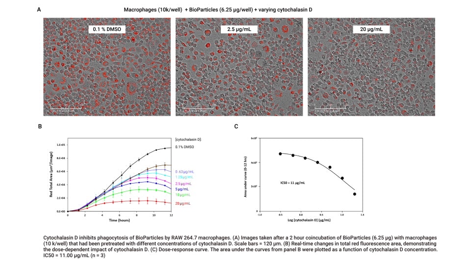
In this example, RAW 264.7 cells are treated with different concentrations of cytochalasin D, an inhibitor of phagocytosis. Compared to the DMSO control, cytochalasin D inhibits phagocytosis in a dose-dependent manner. Plotting the area under the real-time curves as a function of the drug concentration yields a dose-response curve with an IC50 of 11 µg/mL. Easily screen and characterize compounds that modulate phagocytosis with xCELLigence RTCA eSight.


Resources

Real-time visualization and quantitative analysis of macrophage phagocytosis

Using a model system consisting of macrophages and E. coli cells conjugated to a pH-sensitive fluorescent dye, this application note shows the utility of the Agilent xCELLigence RTCA eSight real-time imaging platform for monitoring phagocytosis.

Read app note

New Developments in Cell Health, Viability, and Function

In one instrument, capture sensitive changes in cell number, movement, morphology, and attachment by simultaneous impedance monitoring and live cell imaging. Download the xCELLigence RTCA eSight Live Cell Analysis Brochure to learn more.

Download brochure

Request a Demo

Interested in learning more? Request a demo by clicking on the link below.

Request a Demo
×

Contact Us