- Products
- Chromatography
- Mass Spectrometry
- Certified Pre-Owned Instruments
- Spectroscopy
- Capillary Electrophoresis
- Chromatography & Spectroscopy Lab Supplies
- Instrument Repair
- Sample Preparation
- Chemical Standards
Analytical Instruments & Supplies
- Cell Analysis
- Automated Electrophoresis
- Microarray Solutions
- Mutagenesis & Cloning
- Next Generation Sequencing
- Research Flow Cytometry
- PCR/Real-Time PCR (qPCR)
- CRISPR/Cas9
- Microscopes and Microplate Instrumentation
- Oligo Pools & Oligo GMP Manufacturing
Life Science
- Immunohistochemistry
- Companion Diagnostics
- Digital Pathology
- Hematoxylin & Eosin
- Special Stains
- In Situ Hybridization
- Clinical Flow Cytometry
- Specific Proteins
- Clinical Microplate Instrumentation
Clinical & Diagnostic Testing
- Lab Management
- Lab Consulting
- Software & Informatics
- Genomics Informatics
- Microplates
- Chromatography & Spectroscopy Lab Supplies
Lab Management & Consulting
Lab Software
Lab Supplies
- Dissolution
- Automated Liquid Handling
- Vacuum Technologies
- Leak Detection
Dissolution Testing
Lab Automation
Vacuum & Leak Detection
- Applications & Industries
- Academia
- Biopharma/Pharma
- Cancer Research
- Cannabis & Hemp Testing
- Cell Analysis
- Clinical Diagnostics
- Clinical Research
- Companion Diagnostics
- Infectious Disease
- Energy & Chemicals
- Environmental
- Food & Beverage Testing
- Genomics
- Materials Testing & Research
- Omics
- Pathology
- Security, Defense & First Response
- Vacuum Solutions
- Training & Events
- Agilent University
Mass spectrometry, chromatography, spectroscopy, software, dissolution, sample handling and vacuum technologies courses
- Pathology Education
On-demand continuing education
- Dako Academy
Instrument training and workshops
- Services
-

- Maintenance & Repair
Service Plans, On Demand Repair, Preventive Maintenance, and Service Center Repair
- Lab Operations Management
Software to manage instrument access, sample processing, inventories, and more
- Compliance Services
Instrument/software qualifications, consulting, and data integrity validations
- Instrument Training & Method Services
Learn essential lab skills and enhance your workflows
- Lab & Instrument Relocation Services
Instrument & equipment deinstallation, transportation, and reinstallation
Lab Management Services

- Lab Business Intelligence
CrossLab Connect services use laboratory data to improve control and decision-making
- Lab Enterprise Services
Advance lab operations with lab-wide services, asset management, relocation
- CrossLab Start Up
Shorten the time it takes to start seeing the full value of your instrument investment
- Agilent Community
- Financial Solutions
- Agilent University
- Instrument Trade-In & BuyBack
Other Services Header1
Other Services Header2
- Lab Solution Deployment Services
- Instrument & Solution Services
- Training & Application Services
- Workflow & Connectivity Services
- Oligonucleotide GMP Manufacturing
Pathology Services
Nucleic Acid Therapeutics
- Advance Exchange Service
- Repair Support Services & Spare Parts
- Support Services, Agreements & Training
- Technology Refresh & Upgrade
- Leak Detector Services
Vacuum Product & Leak Detector Services
- Support & Resources
- Agilent Community
- Instrument Support Resources
- Columns, Supplies, & Standards
- Contact Support
- See All Technical Support
Technical Support
- Financial Solutions
- Flexible Spend Plan
- eProcurement
- eCommerce Guides
Purchase & Order Support
- Application Notes
- Technical Overviews
- User Manuals
- Life Sciences Publication Database
- Electronic Instructions for Use (eIFU)
- Safety Data Sheets
- Technical Data Sheets
- Site Prep Checklist
- Brochures
- Catalogs
- Videos
Literature & Videos
- Solution Insights
- ICP-MS Journal
- Certificate of Analysis
- Certificate of Conformance
- Certificate of Performance
- ISO Certificates
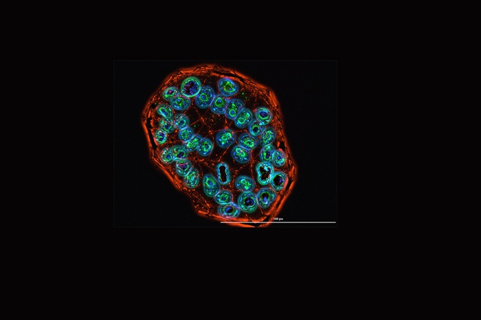
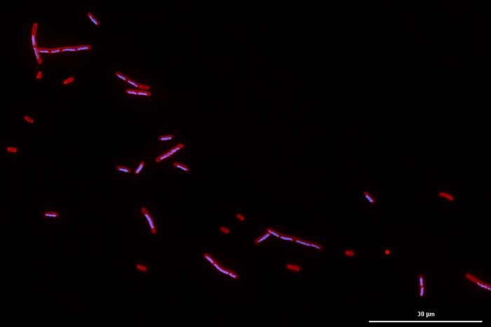
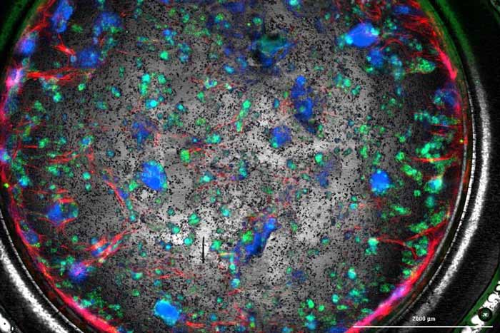
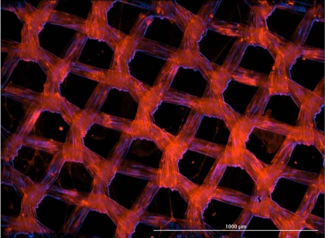
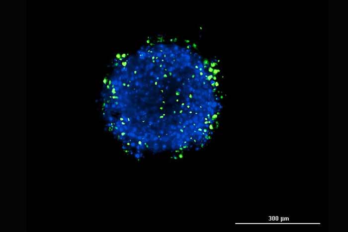
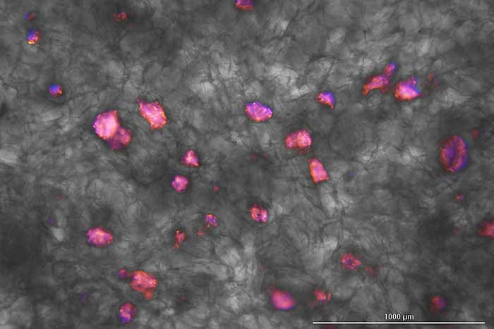
, extracellular polymeric substance (EPS) proteins (red), and EPS polysaccharides (blue). Image shown is a single slice of an image captured on day 8 of development, at 60x confocal with Cytation C10 confocal imaging reader")

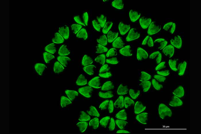
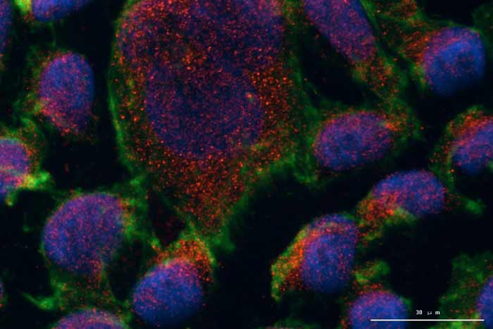
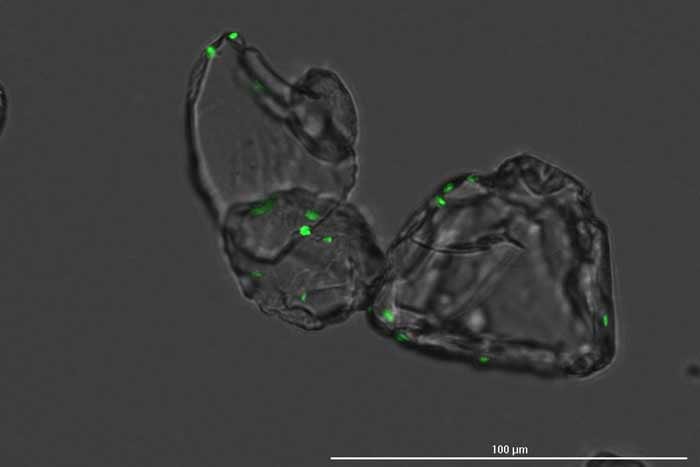
, CF633-conjugated goat anti-rabbit). Captured at 60x water immersion with Cytation C10 confocal imaging reader.")
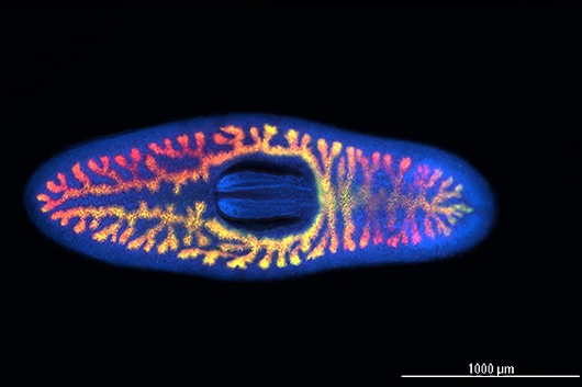
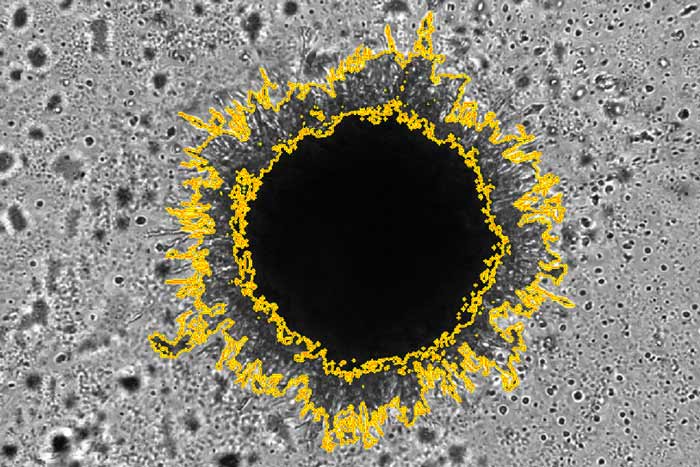
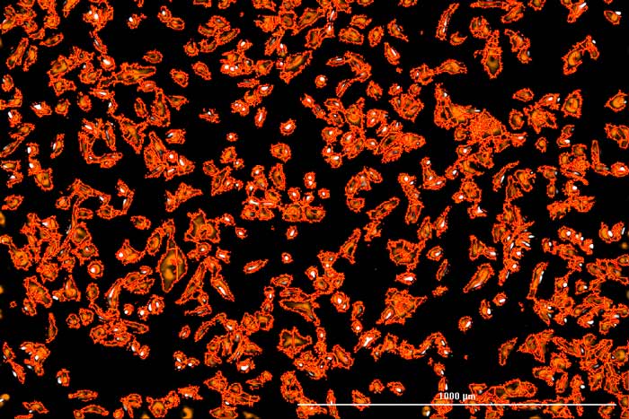
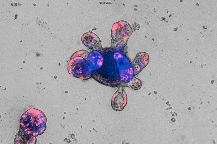
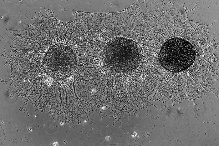
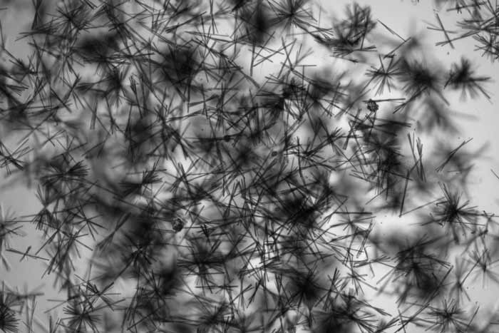
, at 10x brightfield, of a Dictydium cancellatum, a myxomycete of the order Liceales. Pictured is the sporotheca, the bulbous container at the head of the organism that produces and contains the spore mass. Captured at 10x brightfield with Cytation 5 cell imaging multimode reader.")

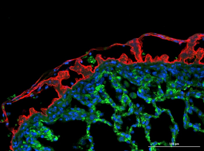
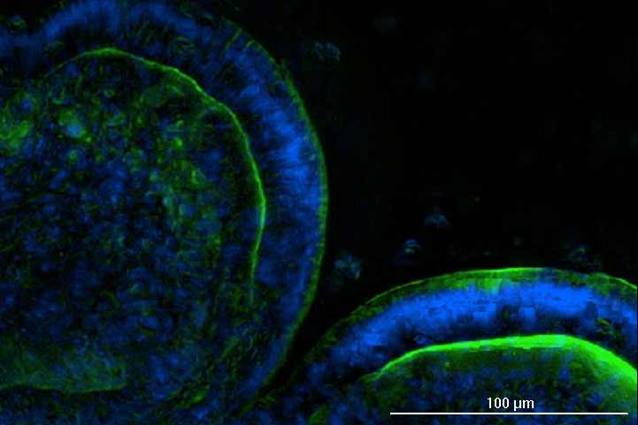
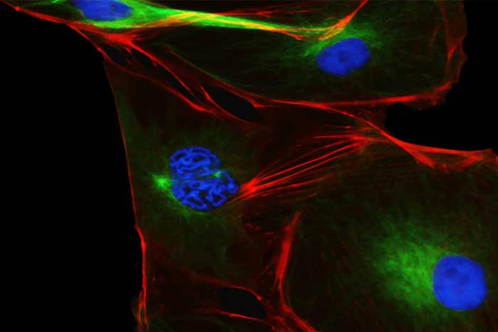
 and counterstained with Alexa Fluor 488-conjugation phalloidin (green) and Hoechst 34580 (blue). Imaged at 60x water immersion. Image captured at 100 µm depth.")
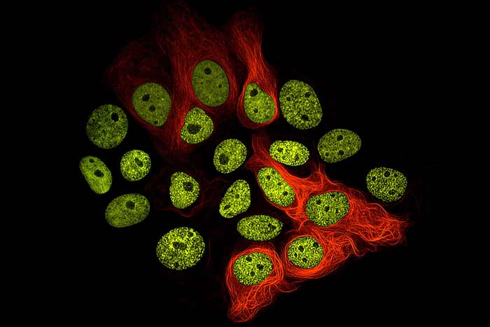
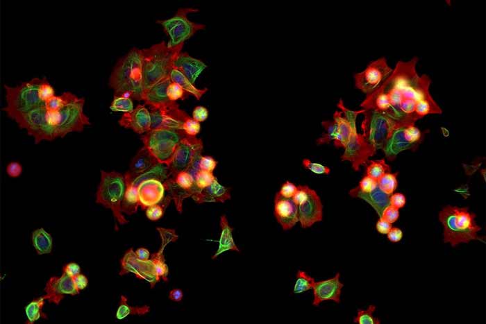
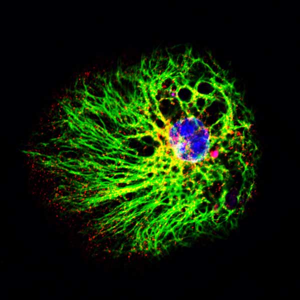
, tubulin (green), nuclei (blue), mitochondria (orange). Widefield 2x2 montage captured at 40x with Agilent BioTek Cytation C10 confocal imaging reader.")


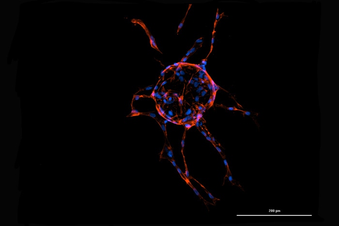
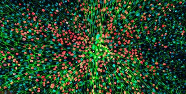
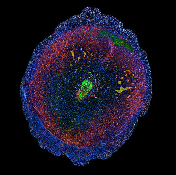
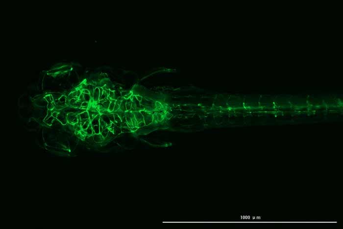
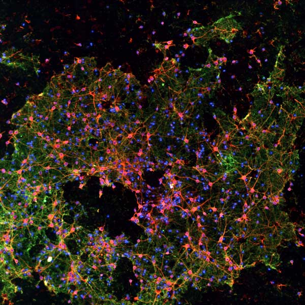
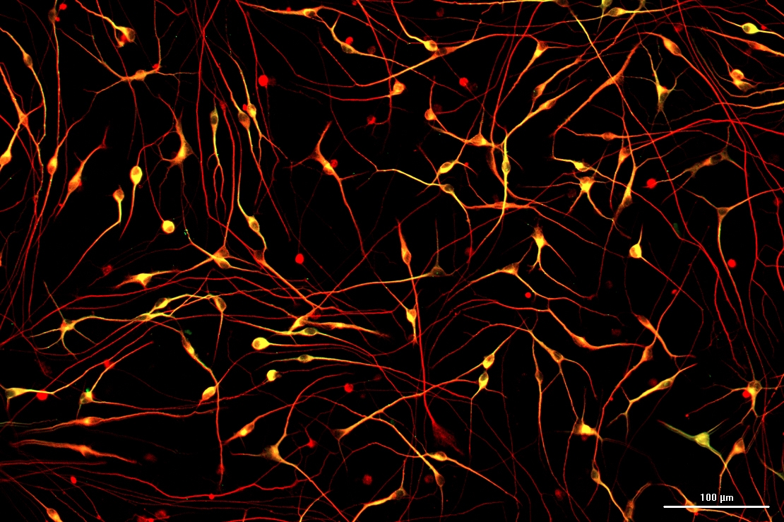
, tyrosine hydroxylase (red) and DAPI (blue). Image captured with Lionheart FX. Summited for the 2021 Imaging contest by Hsueh Fu Wu, University of Georgia")
 from eBioscience and nuclei co-staining using DAPI. The image was taken at a 20x magnification. Submitted for the 2021 Imaging contest by Karina Cereceda of Fundación Arturo López Pérez. Image captured with Cytation 5.")
 taken as a different way to look at cells. Image taken at 40X magnification. Image captured with: Cytation 5. Submitted by Alia Mallah, University of Texas at San Antonio.")
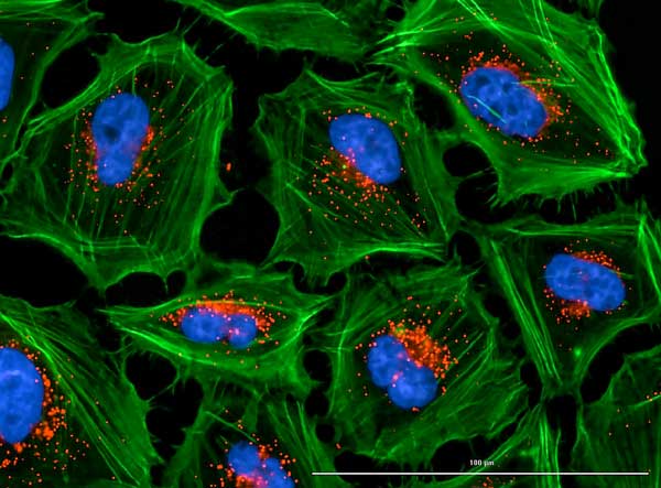
 with the lysosomes (Texas Red channel, anti-LAMP2) which accumulate inside the cells. Image captured with: Lionheart FX by Cristina Andreani, University of Cincinnati")
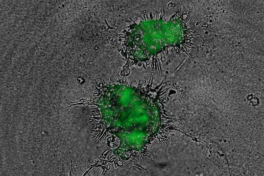
 being targeted and killed by primary human natural killer cell (in red) following treatment with a pro-immunomodulatory agent. Live cell imaging was performed using a Cytation 5 at 10x using two channel fluorescence and brightfield imaging. Image captured with: Cytation 5 by Catherine Mills, Medical University of South Carolina")
, O-GlcNAcylation (red), mitochondria (green), and DAPI. Image was taken at a 10x magnification with automatic image processing. Image captured with: Lionheart FX by Chia-Wei Huang, University of Georgia")
 and fibrin stain (647); moreover, images were taken at 4X. Image captured with: Cytation 5")
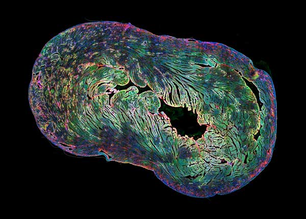
 and H-ATPase (green) in kidney sections of mice. We can observe the cellular conformation of renal tubules and cell nuclei are stained with DAPI (Blue). Image was taken at a 20x magnification. Image captured with: Cytation 1")


, TUJ1 (red), DAPI (blue). Image was taken using a Lionheart FX (manual mode) at a 4X magnification.")

, TUJ1 (green), and DAPI (blue). Image was taken using a Lionheart FX (manual mode) at a 4X magnification.")
, and different phases of cell cycle can be found in this picture")
 with z-stacking every 25 um")

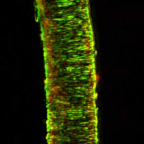
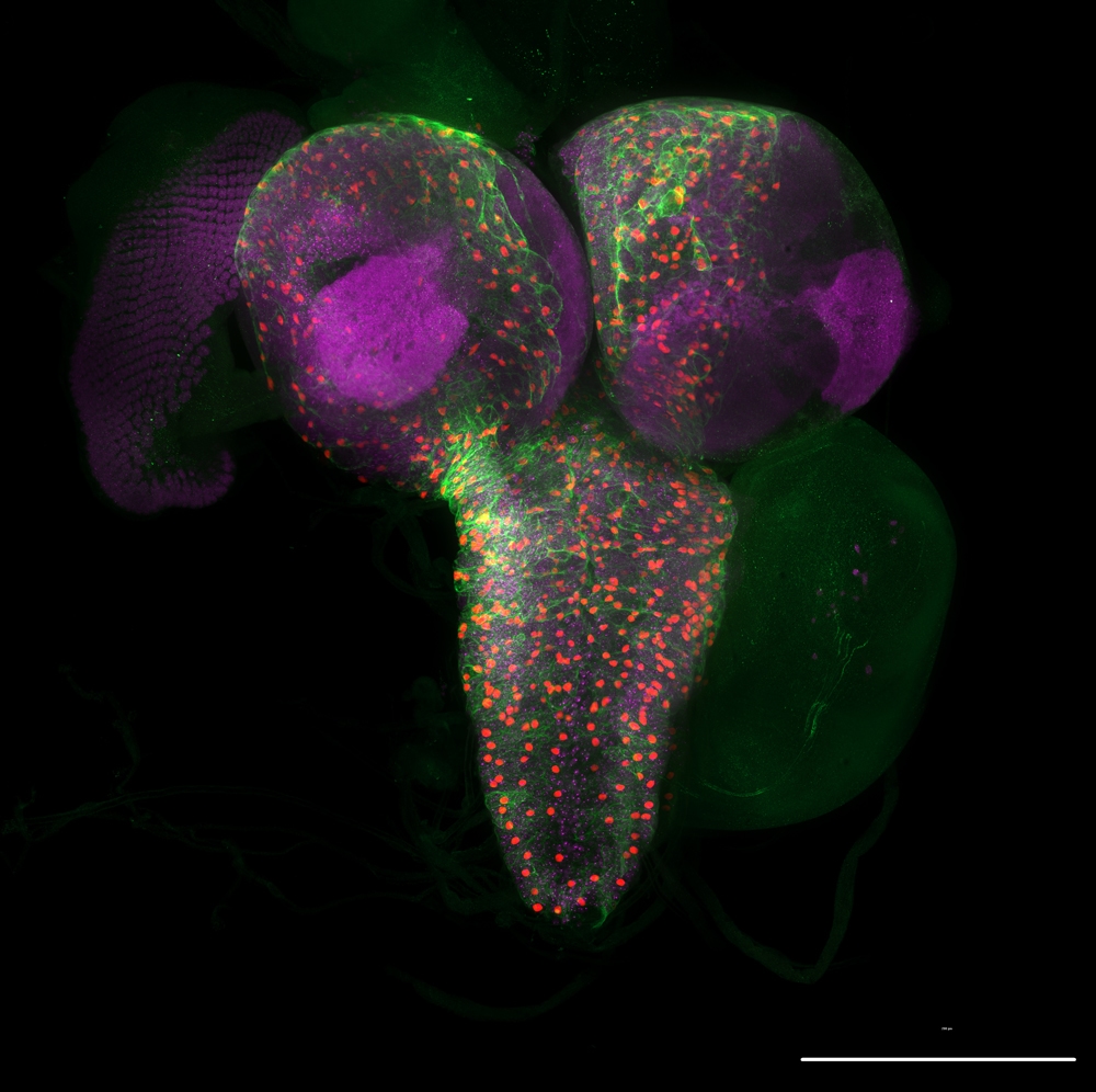
 and neurofilament (Alexa Fluor 647, red) (10X objective).")
 and lysosome GFP (red). Images were acquired using 60X objective. Image shows Z stack projection of maximum fluorescence intensity.")

, and Somatostatin (Alexa Fluor® 488), with DAPI stained nuclei, of porcine islet, imaging at 20x.")
 adipocyte cells stained with BODIPY and Hoechst 33342.")
.")
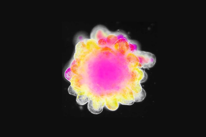
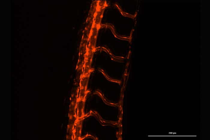
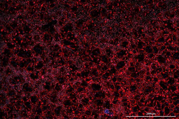
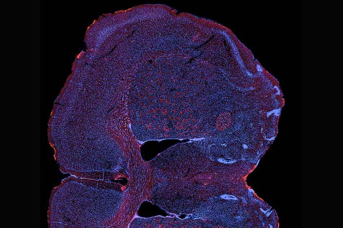
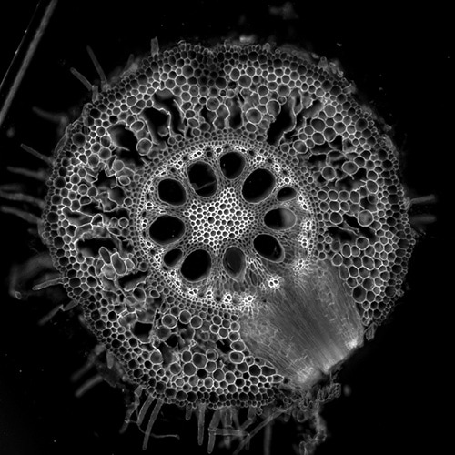
. White/pink color is autofluorescence that indicates vascular cells with enhanced wall thickening. The image was captured at 10x and with brightfield and DAPI, FITC and Rhodamine filters. Captured by Lionheart FX. Submitted for the 2019 Imaging Perspectives contest by Elison Blancaflor, Noble Research Institute LLC.")
, MitoTracker (red) and DAPI (blue). Captured by Cytation 3. Submitted for the 2019 Imaging Perspectives contest by Ibrahim Halil Demirsoy, Luigi Vanvitelli Campania University.")